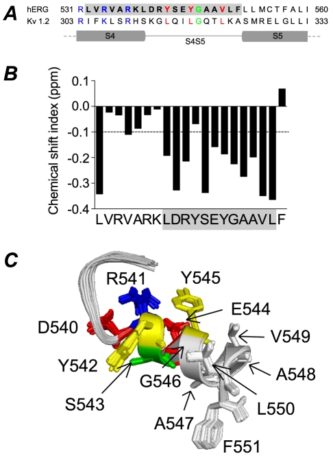
Figure 1. Sequence alignment and structure of hERG S4–S5 linker.
A. Sequence alignment of hERG and Kv1.2 for the distal S4, S4–S5 linker and proximal S5 domains. The leucine residues (red) of Kv1.2 S4–S5 linker correspond to tyrosine and valine residues in hERG. Glycine residue (green) in both channels is also conserved. B. Chemical shift index (CSI) plot for NMR structure of hERG S4–S5. CSI values less than −0.1 ppm are indicative of α-helical structure. C. 20 lowest energy structures for hERG S4–S5 with side chains colour coded according to physiochemical properties (basic: blue, acidic: red, polar: green, aromatic: yellow, hydrophobic: grey).

