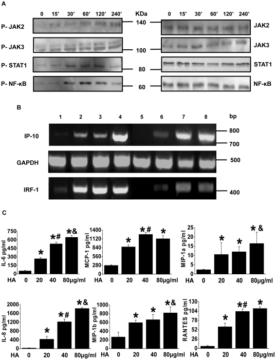
Figure 2. Impact of H5N1 HA on JAK/STAT and NF-κB signalling in the challenged pulmonary epithelial cells.
(A) Detection of phosphorylated/nonphosphorylated JAK2, JAK3, STAT1 and NF-κB. Using specific antibodies, western blotting was performed in the A549 cells treated with the HA (40 µg/ml) for the indicated time periods. Representative blots from 3 replicates are shown. (B) The mRNA expression of IP-10 and IRF-1 on the HA-treated A549 cells. A549 cells were treated with the HA (40 µg/ml) for 1–4 h (Lane 1, 0 h; Lane 2, 1 h; Lane 3, 2 h; Lane 4, 4 h) or with the HA for 1 h at the indicated doses (Lane 5, control; Lane 6, 20 µg/ml; Lane 7, 40 µg/ml; Lane 8, 80 µg/ml) and then subjected to RT-PCR analysis for IP-10 and IRF-1. Representative gels from 3 replicates are shown. (C) Levels of IL-6, IL-8, MCP-1, MIP-1α, MIP-1β and RANTES in the supernatant of A549 cells treated with the indicated doses of HA for 12 h. *P<0.05 vs. control group; # P<0.05 vs. 20 µg/ml HA group; & P<0.05 vs. 40 µg/ml HA group.

