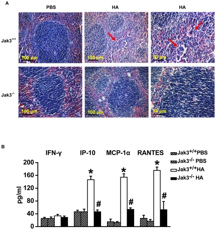
Figure 5. Pathological examination of splenic tissues in the Jak3−/− and Jak3+/+ mice following exposure to HA.
(A) Haematoxylin/eosin (H&E) staining of paraffin sections of splenic tissues from the Jak3+/+ and Jak3−/− mice intratracheally administered with PBS or HA for 72 h. Arrows show the necrosis of lymphocytes. (B) The cytokines/chemokines (IFN-γ, IP-10, MCP-1α and RANTES) that were released from the splenocytes of either Jak3+/+ or Jak3−/− mice pretreated with HA or PBS as described above. The measurement of the concentration of the cytokines/chemokines by Liquidchip assay. *P<0.05 vs. Jak3+/+ PBS group; # P<0.05 vs. Jak3+/+ HA group.

