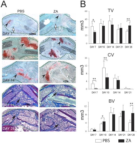
Figure 1. Effects of Zoledronate on the course of tibial fracture repair.
(A) Sections through the calluses (green dashed-lines) of PBS- (left) and zoledronate- (right) treated mice were stained with Safranin-O/Fast Green (days 7, 10 and 14 post-fracture) to detect cartilage (arrowsheads) and Trichrome (days 21 and 28 post-fracture) to detect bone (arrows). White arrows indicate the fracture site. Scale bar = 500 µm. (B) Histomorphometric analyses of total callus volume (Top), total cartilage volume (CV, middle) and total bone volume (BV, bottom) on PBS treated and ZA treated-mice at days 7, 10, 14, 21 and 28 post-fracture (n = 6 per group). *p<0.05, **<0.01. Bars represent mean±s.d.

