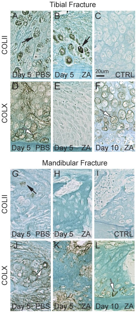
Figure 3. Effects of Zoledronate on collagen types II and X expression during tibial and mandibular fracture repair.
Immunostaining of collagen type II (A–B, G–H, black arrows) and collagen type X (D–F, J–L, white arrows) near the periosteum within tibial (top) and mandibular (bottom) fracture calluses of PBS (left) and zoledronate (middle and right) treated mice. No staining is observed in negative controls (C and I).

