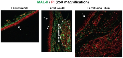
Figure 2. α2–3 linked glycan distribution in ferret respiratory tract.
MAL-II lectin (green) was used to stain ferret cranial, caudal and lung hilar regions. As seen from the images, MAL-II stained the submucosal glands, the underlying mucosa and some goblet cells (marked as *) in the caudal region. There was no staining of the lung hilar region indicating an absence of α2–3 glycans. The nuclei were stained with PI (red). The apical surface is marked with a white arrow.

