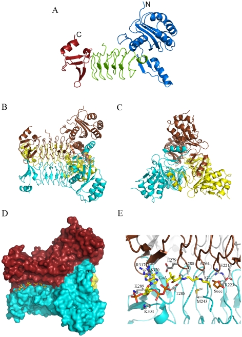
Figure 3. The structure of P. aeruginosa DapD.
A. Cartoon of the subunit of tetrahydrodipicolinate N-succinyltransferase (DapD). The N-terminal domain is shown in blue, the central domain in green and the C-terminal domain in red. B & C: Views of the trimer of DapD. The three subunits are coloured blue, brown and yellow. D. Surface illustration of the trimer of the DapD-coenzyme A-succinate complex. The substrate binding grooves are formed between the left handed β-helix domains from adjacent subunits (blue and brown, respectively) of the trimer. Bound co-enzyme A and succinate are shown as stick models in yellow. E. Interactions of DapD with the bound coenzyme-A and succinate. The two subunits contributing to this active site are shown in blue and brown, respectively. Coenzyme-A is shown in yellow sticks and succinate in orange, while the amino acid side chains involved in the interaction are depicted in light gray.

