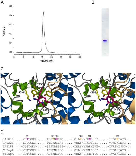
Figure 7. The structure of P. aeruginosa DapA.
A. Size exclusion chromatography elution profile of PaDapA (PA1010) indicating that a single species exists in solution. Based on the calibration curve (insert) the calculated molecular mass is 60 kDa. B. PaDapA (PA1010) purified sample analyzed in native polyacrylamide gel electrophoresis indicating a single species. C. Stereo view of the active site of PaDapA located in the center of the α/β barrel. Amino acid side chains forming the active site are indicated as stick models. Residues conserved in the three homologues PA1010, PA0223 and PA4188 are shown in yellow, while the variable positions Thr44, Arg138 and Lys109 are indicated in purple. D. Sequence conservation in DapA enzymes from Escherichia coli, Bacillus anthracis, Pseudomonas aeruginosa and the proposed DapA paralogues in the PAO1 genome PA0223 and PA4188. The active site residues in PaDapA (PA1010) are indicated with yellow or purple colour as in C.

