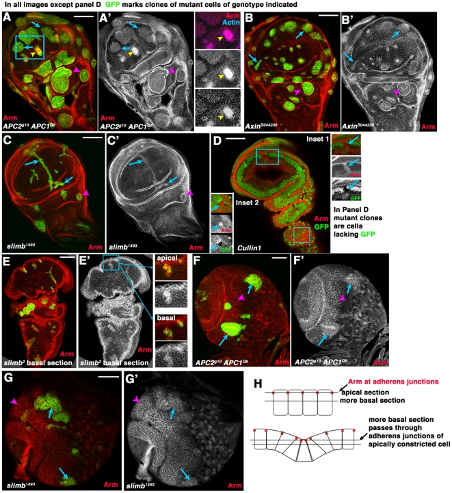
Figure 4. Arm accumulates to similar levels in wing imaginal disc cells mutant for different destruction complex or SCF proteins.
A–E. 3rd instar wing imaginal discs. F,G. 3rd instar larval brains. In all cases except D clones of mutant cells of the indicated genotype were induced using the MARCM method [42] and homozygous mutant cells are marked by the presence of GFP. In D, homozygous mutant cells have lost GFP. A–C. Arrows, cells in the wing pouch mutant for both APCs (A), Axin (B) or slimb (C) all accumulate modestly elevated levels of Arm. Arrowheads, mutant cells in regions surrounding the wing pouch segregate and form cysts. A. Inset. Double mutant cells appearing to accumulate more elevated Arm levels are sectioned through the top of apically constricted cells, as demonstrated by their constricted apical ends and elevated actin accumulation in that plane of focus. D. We obtained very few and small clones mutant for Cullin1, which are marked by the lack of GFP—they accumulated elevated levels of Arm (Insets, mutant cells shown by arrows). E. slimb mutant cells, marked by GFP. Insets show an apical and more basal section through the same clone. Arm accumulation appears very high in apical section, but more basal section reveals more modest accumulation. Apical sections pass through the adherens junctions of mutant cells, which have apically constricted (diagrammed in H), creating the impression of more highly elevated Arm levels. F,G. Arrows, cells in the medulla mutant for both APCs (F) or slimb (G) accumulate modestly elevated levels of Arm. Arrowheads, wild-type cells showing normal levels of accumulation in this tissue. H. Diagrams illustrating changes in morphology in mutant clones and resultant effect on plane of focus in wild-type cells and mutant neighbors. Scale bars = 50 µm.

