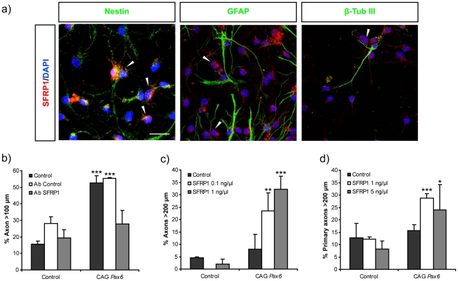
Figure 2. SFRP1 secreted by cortical cells in vitro is responsible for the axonal growth observed in Pax6 over-expressing neurons.
a) Immunofluorescence shows expression of SFRP1 (red) in differentiating, NSCs cultured for 5 days. Cells were double labeled with anti-nestin, GFAP and β-tubulin III. SFRP1 expression is found in the cytoplasma of Nestin-positive cells or in some with a low GFAP expression (arrowheads). No SFRP1 expression was found in cell with high GFAP levels or in β-tubulin III-positive cells. Bar represents 20 µm. b) The graphs show the percent of axons longer than 100 µm upon Pax6 overexpression in NSCs when cultured in the presence or absence of control or anti-SFRP1 antibodies. Note that antibody against SFRP1 blocks axonal growth stimulated by the ectopic expression of Pax6. c, d) Response to increasing concentrations of purified recombinant SFRP1 in neurons derived from NSC c) or primary cortical neurons (d). NSCs (c) and primary neurons (d) were transfected with control empty CAG and CAG-Pax6 vectors and stimulated with different concentrations of SFRP1.Addition of recombinant SFRP1 only stimulates the growth and elongation of axons of neurons ectopically expressing Pax6. Data are expressed as the mean ± SD. (*) p<0.05; (**) p<0.01; (***) p<0.001. Number of axons per condition >50 (n = 3).

