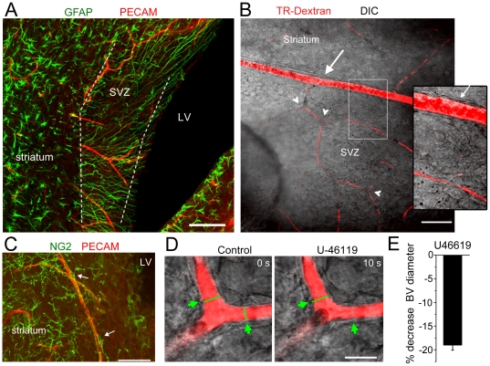
Figure 1. U-46119 constricts SVZ capillaries in acute slices.
(A) Z-stack projection of GFAP staining (green, mature and SVZ astrocytes) and PECAM (red, blood vessels) in a horizontal section. The dashed lines encompass the SVZ. LV: lateral ventricle. (B) Texas Red (TR)-Dextran-filled vessels coursing through the SVZ in a live sagittal section. An arteriole (arrow) branches into capillaries. Note the presence of capillary branchpoints (arrowheads). Inset: zoom of the region delineated by the white rectangle in B. The white arrow points to the smooth muscle cells around the arteriole. (C) Z-stack projection of NG2 (green) and PECAM (red) immunofluorescence in a coronal section. NG2 cells on capillaries are pericytes (arrows). (D) Image of the capillary before (control) and during U-46119 (100 nM) application. The capillary was loaded with TR-Dextran through cardiac perfusion prior to slicing. The green arrows indicate the sites of constriction. (E) Mean % change in blood vessel diameters during and after U-46119 applications. Scale bars: 30 (A), 50 (B), 40 (C), and 15 µm (D).

