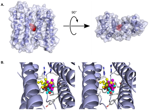
Figure 3. Diverse general anesthetics utilize a common binding site in apoferritin.
A) Orthogonal views of the apoferritin dimer, shown as a partially transparent molecular surface encasing a cartoon representation of the backbone. The anesthetic binding site is marked by thiopental, which is shown in a red space-filling representation. B) Stereo view showing a close-up of the anesthetic binding site, in an orientation similar to that seen in the left half of the upper panel. Four different general anesthetics are shown in ball-and-stick representations: Thiopental (yellow); propofol (magenta); isoflurane (cyan); and halothane (orange). The protein backbone is shown in blue, while selected protein side chains are shown in light gray. All four compounds, despite belonging to different chemotypes, utilize the same binding cavity, in which their positions overlap extensively.

