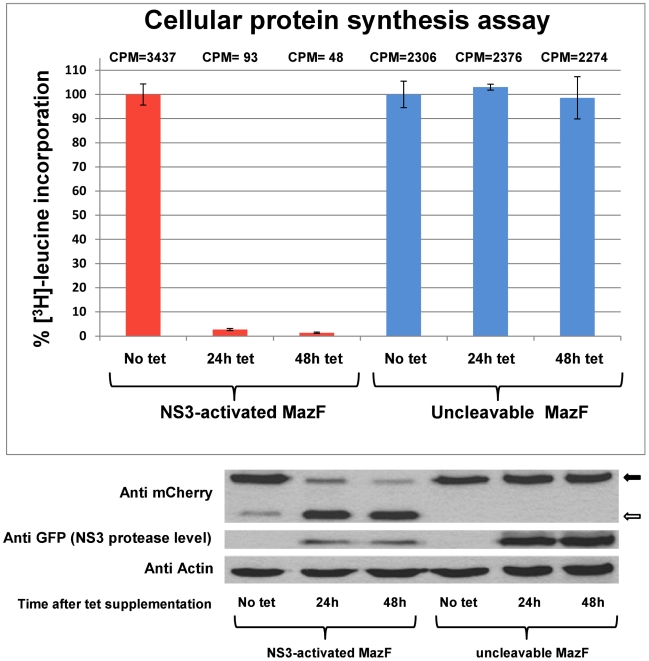
Figure 4. Inhibition of de-novo protein synthesis by NS3-activated MazF based zymoxin in NS3-expressing cells.
1×10⁵ Tet-NS3/activated MazF or Tet-NS3/uncleavable MazF cells were seeded per well in 24-wells plate. 24 or 48 h later, cells were supplemented with tetracycline to a final concentration of 1000 ng/ml, or left untreated (48 h tet, 24 h tet and no tet, respectively). 72 h after seeding, levels of de-novo protein synthesis were determined by [3H]-leucine incorporation assay, as described in “materials and methods”. Results are expressed as percent of the value obtained for cells which were not induced to express the NS3 protease (No tet). Each bar represents the mean ± SD of a set of data determined in triplicates. Numbers above each bar represent mean counts per minute (CPM) values for 7 micrograms total protein samples (upper panel). 30 micrograms of total protein from lysates of the described cells were analyzed by immunoblotting with mouse anti-mCherry (for detection of the zymoxin), mouse anti-GFP (for the detection of EGFP-NS3) and mouse anti actin antibodies (loading control) followed by HRP-conjugated secondary antibodies and ECL development. Solid arrow: full length zymoxin. Hollow arrow: N' terminal portion of NS3-cleaved zymoxin (lower panel).

