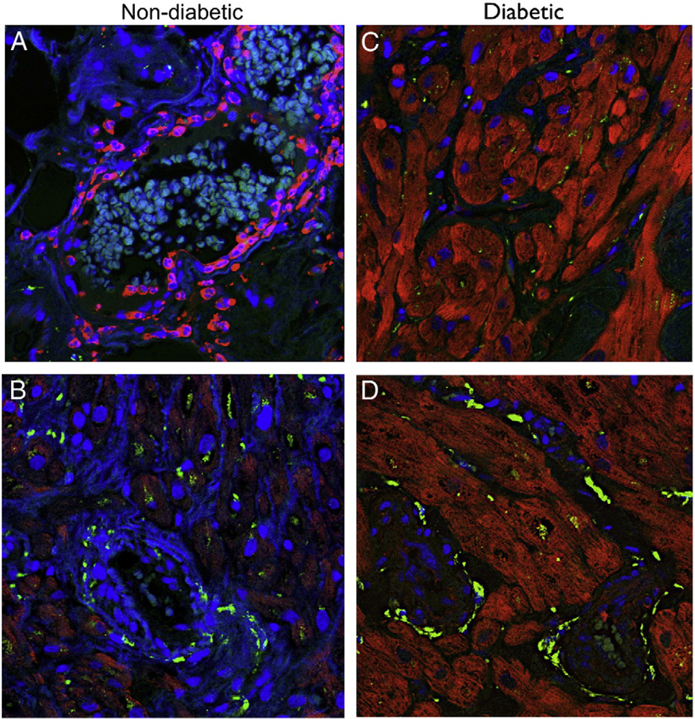
Fig. 10.
Immunofluorescence images. Representative slides of atrial tissue from non-diabetic and diabetic patients. Sections were co-stained with 4′,6′-diamidino-2-phenylindole (DAPI, blue) to demarcate the nucleus and Y5 receptors. Magnification for all images is 40×. There is increased staining for Y5 receptors in diabetic tissue cardiomyocytes (C and D) as compared to non-diabetic tissue where the localization of Y5 receptors is around blood vessel. (A and B).

