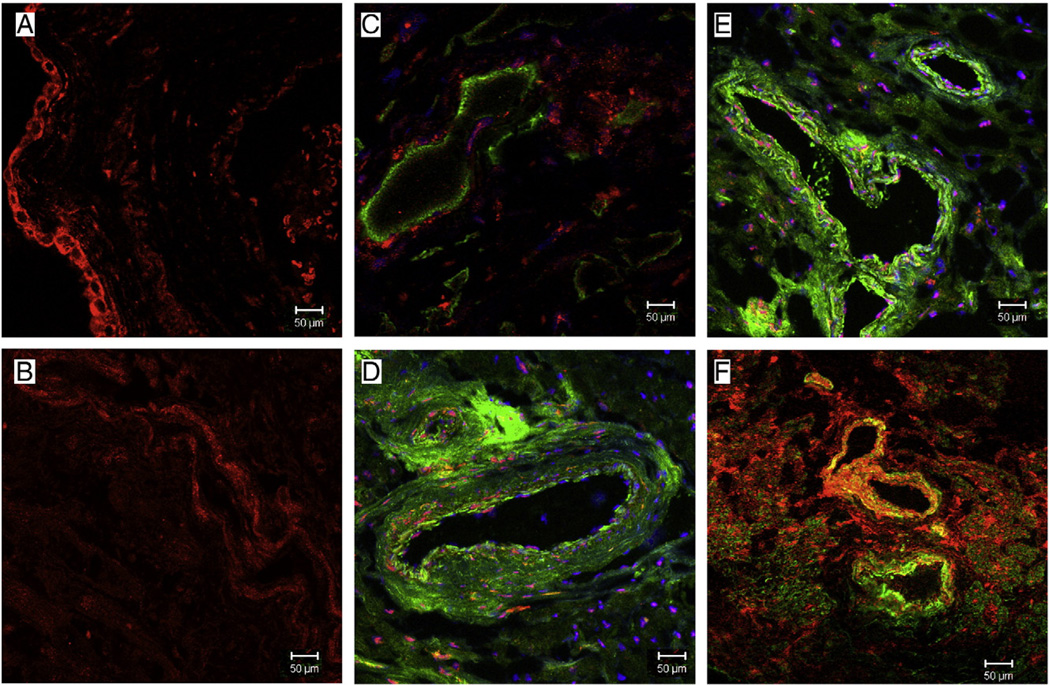
Fig. 7.
Representative immunofluorescence staining for Y1 (red) in sections co-stained with the vascular smooth muscle marker -smooth muscle actin (green) in atrial tissue from Non-diabetic and Diabetic patients. (A and B) There is staining of Y1 receptor in the endothelium of non-diabetic tissue, while the staining is decreased in the endothelium of diabetic tissue. (D and F) There are increased staining of Y1 receptor around the arteries in the atrial tissue of diabetic patients as compared to non-diabetic patients (C and E).

