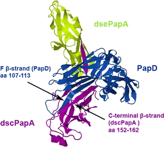
Fig. 3.

Overall 3D structure of the PapD/PapA/PapA complex (PDB ID 2UY6): PapD (blue), donor–strand complemented (dsc) PapA (purple), and donor–strand exchanged (dce) PapA (green) are shown in ribbon representation with β-stands as arrows and α-helixes indicated as cylinders. The F-β-stand of PapD (amino acids 107–113) is indicated by an arrow, as is the C-terminal β-stand of dscPapA (amino acids 152–162)
