
Figure 4.
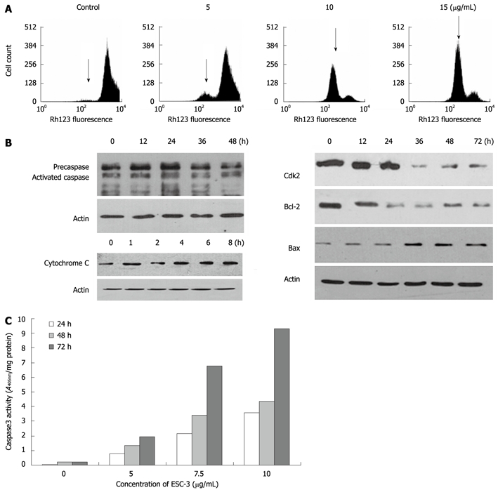
Apoptosis induced by ESC-3 through the mitochondria-dependent pathway. A: Effect of ESC-3 on the ΔΨm of cholangiocarcinoma cells. The increase in Rh123 hypofluorescence indicates a reduction in ΔΨm, which is shown with arrows; B: Expression of cytochrome C, caspase-3, CDK2, Bax, and Bcl-2 in Mz-ChA-1 cells treated with 10 μg/mL ESC-3 for different periods of time; C: Effect of ESC-3 on the activation of caspase-3 activity in Mz-ChA-1 cells. The cells were treated with 0 μg/mL, 5 μg/mL, 7.5 μg/mL and 12.5 μg/mL ESC-3, and caspase-3 activity was analyzed after 24 h, 48 h or 72 h.