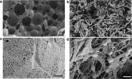
Figure 1. Electron microscopic analysis.
Significant differences in terms of micro-nano structure were observed between HA and HA-Col scaffolds (a–c and b–d respectively). At low magnification, HA foams show round-shaped interconnected pores, with black spots representing interconnections between neighbor pores (a). Ha-Col sponge, on the other side, shows a heterogeneous fibrous structure (b). At higher magnification, HA grains clearly form a substrate for cell adhesion, with black spots representing microporosity (c). Ha-Col scaffold highlights cells grasped to the collagen nanofibers (d). Bars: 200 μm, 500 nm respectively.

