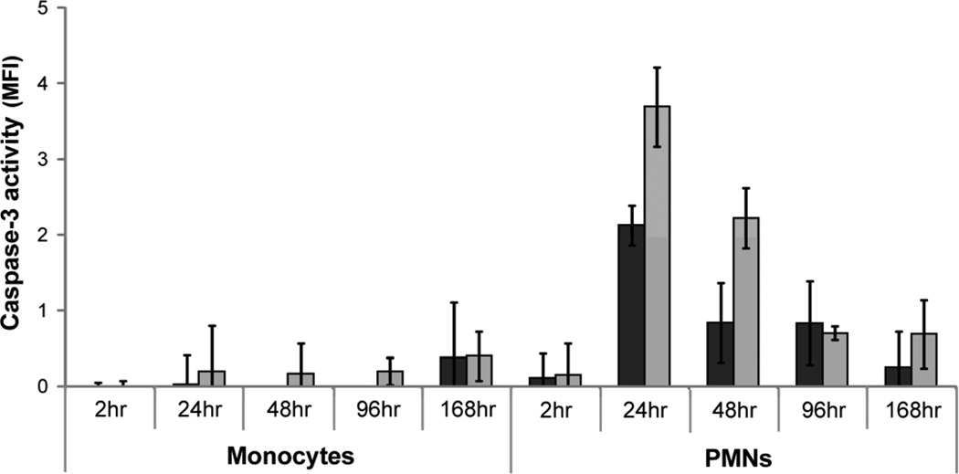
Figure 8.
Levels of secondary necrosis present in monocyte and PMN cultures as determined by measuring the activity of caspase-3 in the supernatant. Supernatant from cells exposed to TCPS ( ) and PEG hydrogel (
 ) substrates for 2, 24, 48, 96, and 168hr were exposed to fluorogenic caspase-3 substrate and the mean fluorescence intensity from the cleaved probe was assessed.

