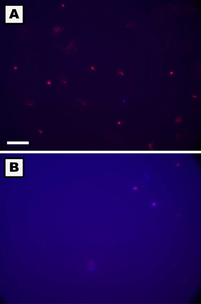
Figure 9.
Photomicrographs of apoptotic and necrotic monocytes on TCPS (A) and PEG hydrogel (B) surfaces at 24hr. Apoptosis detection was based on the translocation of phosphatidylserine from the inner to outer membrane of the plasma membrane in apoptotic cells (Blue). Primary and secondary necrosis was detected using the nucleic acid-binding propidium iodide dye which cannot penetrate the membranes of live or early apoptotic cells (Red). Live cells demonstrated no or a very low level of fluorescence. The PEG hydrogel created blue background fluorescence. The white reference line represents 75µm.

