
Figure 5. p150Sal2 binds in vitro to fragments of the p21Cip1/Waf1 promoter containing GC boxes.
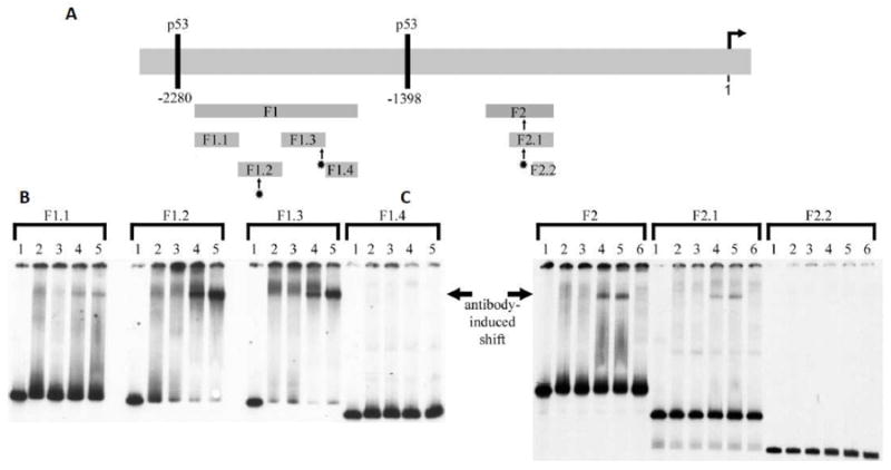
A – Schematic of the ~ 2.6 kb promoter showing fragments F1 and F2 and subfragments generated by Xho and HindIII analyzed by EMSA. B and C – EMSA results showing binding of p150Sal2 to subfragments F1.2, F1.3 and F2.1 as indicated (-). Radioactively labeled subfragments were incubated with nuclear extracts of 293 cells and the products analyzed by EMSA. Antibody to p150Sal2 was used to induce supershifts in subfragments F1.2, F1.3 and F2.1. Lanes 1: DNA probe alone. Lanes 2: probe + nuclear extract. Lanes 3: probe + nuclear extract + antibody to Sp1 (control). Lanes 4: probe + nuclear extract + antibody to N-terminus of p150Sal2. Lanes 5: probe + nuclear extract + antibody to C-terminus of p150Sal2. Lanes 6: probe + nuclear extract + antibody buffer control. Antibody-induced shifts in mobility and focusing of bands are indicated (¢¡). Subfragments F1.2, F1.3 and F2.1 contain GC boxes. [See text].