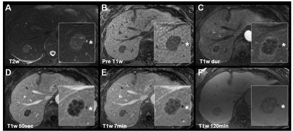
Figure 10.
Metastatic disease imaged with gadoxetic acid in a 73 year old female with known rectal carcinoma. Typical enhancement patterns of a hypo-vascular metastasis can be appreciated: slight hyper-intensity on T2w images, marked hypointensity on T1w pre contrast, peripherally dominant and slow central enhancement leading to the previously described ‘target sign’ with marked hyperintensity on the 120min hepatobiliary phase. A small cyst (*) is also noted, adjacent to the lesion of interest.

