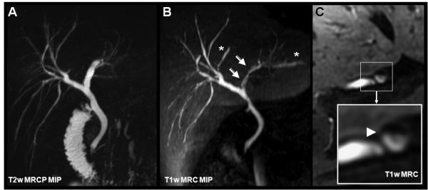
Figure 14.

Gadoxetic acid for T1w MRC in a 35-year-old male with ulcerative colitis. Subtle, alternating strictures and beading are with both T2w MRCP (A) and T1w MRC (B). Note the apparent stenosis in B (open white arrows) that is caused by layering of contrast in the dilated duct (see cross-sectional magnifications in C). Asterisks indicate slightly dilated bile ducts not seen by T2w MRCP due to incomplete coverage by the limited slab thickness of the MRCP.
