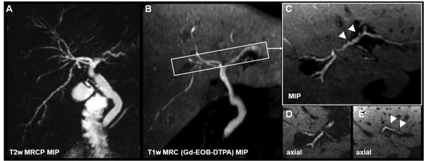
Figure 15.
Beaded appearance of the biliary tract imaged with gadoxetic acid in a 44yo male with known primary sclerosing cholangitis (PSC). Although the high liver signal limits the ability to fully appreciate MIP representations of the bile ducts in the same manner as MRCP, the axial detail allows for identification of biliary duct irregularities PSC with exquisite detail.

