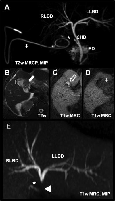
Figure 16.

Bile leak in a 53 year-old after laparoscopic cholecystectomy using gadoxetic acid. (A) T2w MRCP with the cystic duct remnant (*) and a surgical drainage pigtail catheter (‡) placed in the gallbladder fossa. Axial T2w imaging (B) shows postsurgical fluid collection in the fossa (white arrow). 30min after injection of gadoxetic acid, there is contrast agent accumulation in the fossa (white open arrow, C). (D) shows contrast within the pigtail catheter (‡). The T1w MRC MIP in (E) demonstrated an abrupt termination of the contrast column in the common duct superior to the level at which the contrast leaks entirely into the gall bladder fossa and subsequently into the pigtail drainage catheter. This example demonstrates the advantage of the functional information that can be obtained using hepatobiliary agents. CHD = common hepatic duct; PD = pancreatic duct; RLBD and LLBD = right and left biliary duct, respectively.
