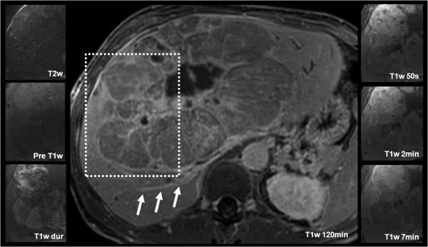
Figure 5.
Findings in a rare case of fibrolamellar HCC evaluated with gadobenate dimeglumine in a 42 year-old male with elevated liver function tests and jaundice. This enormous liver tumor demonstrated heterogeneous pre-contrast T2w and T1w signal properties. The solid components of the tumor showed a similar rapid arterial contrast enhancement and portal venous wash-out as the HCC lesion in figure 5. The radial septae with progressive delayed enhancement (E, F) mirror the fibrous components of this tumor. Also note the contrast in the dilated biliary duct in the posterior right lobe on the 120 minute image (white arrows).

