
Figure 7.
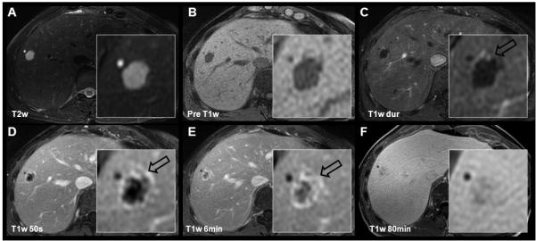
Cavernous hemangioma and simple cyst depicted using gadobenate dimeglumine. 56 year-old female referred to MR with multiple indeterminate lesions and history of uterine cancer. The cyst is easily characterized by its “light bulb” bright appearance on T2w images and lack of enhancement on post-contrast images. The cavernous hemangioma shows slightly lower T2w hyperintensity, and centripetal contrast enhancement (open black arrows). Note near isointensity of the hemangioma on the 80 minute delayed gadobenate dimeglumine images as opposed to relative hypointensity using gadoxetic acid (Figure 6).