
Figure 8.
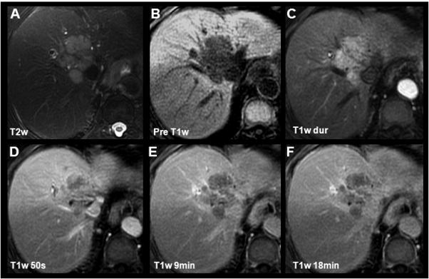
Characterization of a cholangiocarcinoma using gadobenate dimeglumine. In this 80yo female an intrahepatic CC with characteristic pre-contrast imaging findings, rapid T1w contrast enhancement and portal venous washout was depicted. Slight capsular enhancement can be appreciated (D) consistent with either compressed neovasculature or fibrous tissue. Of special note is the isointensity of the CC in the delayed phase (18min, F) compared to the relative hypointensity when imaging with gadoxetic acid (Figure 9).