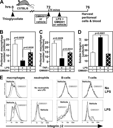
FIGURE 3.
In vivo administration of an MMP/ADAM inhibitor GM6001 prevents LPS-induced macrophage exiting from the peritoneum and release of soluble integrin β2, while increasing β2 surface expression on macrophages. A, mice received thioglycollate and after 3 days (3–5 mice per group) were injected with the metalloprotease inhibitor GM6001 or vehicle 15 min prior to administration of vehicle or 1 μg of LPS, all by intraperitoneal injections. Four hours later, cells from the peritoneal cavity were harvested, counted, and stained for flow cytometric analysis of cell distributions. B, macrophage (F4/80+, Ly6G−) numbers in the peritoneal cavity were determined. C, peritoneal macrophages were collected as shown in A except that vehicle or 0.5 μg of TNF-α was administered 15 min following injection of GM6001. Macrophage (F4/80+, Ly6G−) numbers in the peritoneal cavity were determined. D, peritoneal lavage fluids were analyzed 4 h following injection of PBS, LPS or LPS with GM6001 by αMβ2 ELISA. E, representative FACS histograms of two mice from each group show the mean fluorescence intensity with (gray) or without (black) GM6001 for integrin β2 on macrophages, neutrophils, B-cells, and T-cells in the presence (lower panel) or absence (upper panel) of LPS. Means ± S.E. are shown, and data are representative of three different experiments.

