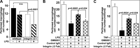
FIGURE 4.
In vivo administration of disrupting integrin β2 antibody 2E6 promotes macrophage exit from the peritoneum prevented by the metalloproteinase inhibitor GM6001. A, mice either integrin β2 +/+ or −/− received an LPS injection as described in Fig. 3A, and then cells were harvested after 2 h to evaluate exiting (n = 4–5/group; n.s. = not significant). B and C, experimental protocol to measure macrophage exiting was the same as in Fig. 3A except that the 15-min incubation with GM6001 or vehicle control also included treatment with 150 μg of 2E6 Fab fragments or hamster isotype IgG control prior to LPS injection. Macrophage cell number was quantitated by FACS analysis (F4/80+, Ly6G−). The numbers of macrophages remaining in the peritoneal cavity with different treatments is shown: B, following LPS administration (n = 3–4/group); C, after TNF-α stimulation (n = 3/group). The data are expressed as the number of macrophages that remain in the peritoneal cavity after 2 or 4 h. Means ± S.E. are shown, and the data are representative of two replicate experiments.

