FIGURE 1.
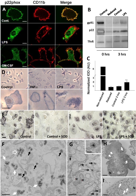
LPS and GM-CSF induce redistribution of cyt b558 from the cell surface to an intracellular membrane compartment in primary macrophages. A, rat primary microglia were treated with 100 ng/ml LPS or 10–20 ng/ml GM-CSF overnight and then processed for immunofluorescence with polyclonal rabbit anti-p22phox antibodies (green, left column) and anti-CD11b mAb (red, middle column). The merged channels are shown in the right column. Bars, 10 μm. B, internalization of cyt b558 subunits in rat BMDMs after stimulation with 100 ng/ml LPS for 3 h was detected by a cell surface biotinylation protocol (NHS-SS-biotin). The bar graph in C represents optical density mean ± S.E. of Western blot bands for p22phox derived from three independently performed experiments. Note that the reduced and “treated” lanes, which appear as separate gel strips in B, were derived from nonadjacent lanes from the same gel. AU, arbitrary units. D, primary microglia were stimulated overnight with 20 ng/ml GM-CSF or 100 ng/ml LPS before stimulation with 100 ng/ml PMA in the presence of NBT. Note the redistribution of the blue reaction product from the cell surface of untreated control cells (delineating the cell periphery) to an intracellular punctate pattern following stimulation. Bars, 10 μm. E, human monocyte-derived macrophages treated or not with 2 μg/ml LPS overnight were stimulated with 100 ng/ml PMA with or without 400 units/ml SOD in the presence of NBT salt. Bars, 10 μm. F–I, CeCl3 cytochemistry on LPS-treated BMDM or Ra2 microglia, which were stimulated with PMA in the presence of CeCl3 for 1 h before processing for EM. F, in BMDM, the electron-dense cerium hydroxide reaction product is deposited in small 100 nm vesicles (black arrows), tubulovesicular elements (open arrow), and large vacuoles (white arrows). G, cerium hydroxide deposition in Ra2 cells was less intense, but the same compartments as above could be identified. Shown here are examples of 100 nm vesicles with clear internal precipitates (see inset for detail). Note that other membranes, including mitochondria (M), are devoid of reaction product. Bars, F and G, 500 nm (inset in G, 100 nm). H, Ra2 microglia; I, BMDM at low magnification. The reaction product is deposited almost exclusively inside both cell types (arrows) with little or no product on the plasma membrane. N, nucleus. Bars, 1 μm.

