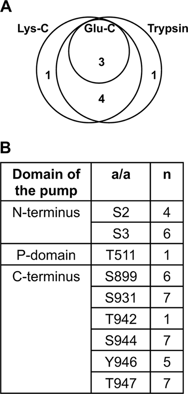
FIGURE 3.

Phosphorylation of recombinant autoinhibited plasma membrane H+-ATPase 2 (AHA2) heterologously expressed in yeast membranes. A, shown is a Euler diagram of the number of phosphosites determined by digesting of AHA2 in a mixture of solubilized membrane proteins with trypsin, Lys-C, and Glu-C. Trypsin and Lys-C were of the same effectiveness and gave one unique site each. B, shown is a summary table of phosphosites in AHA2 determined using complementary digestion by three enzymes, their distribution within the H+-ATPase domains, and number of cases (n) when phosphosites were determined within a total number of 7 samples analyzed. a/a, amino acid. Almost all the phosphosites determined are in the N- and C-terminal ends of the pump.
