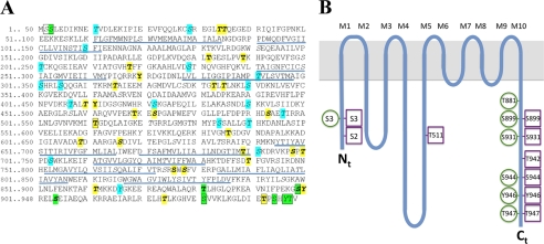
FIGURE 5.
Overview of phosphosites in AHA2. A, shown is the amino acid sequence of AHA2. Green color, phospho-residues identified in AHA2 expressed homologously in planta (this study and previous studies). Boxed, phosphosites identified in recombinant AHA2 expressed heterologously in yeast. Italics (and cyan when not phosphorylated in AHA2 in planta), Phosphosites were predicted by the NetPhosK server. Bold (and yellow when not phosphorylated in AHA2 in planta), phosphosites predicted by the PhosPhAt 3.0 server; underlined, membrane spanning domains in the crystal structure of AHA2 (59). B, shown is a spaghetti model of AHA2 with the phosphosites indicated. Green circles, phosphosites when expressed in planta. Magenta squares, phosphosites when expressed in yeast.

