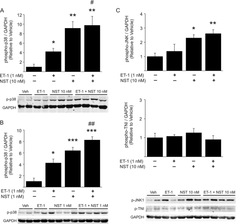
FIGURE 3.
Neuronostatin induces p38 MAPK and JNK phosphorylation in isolated perfused hearts. A–C, hearts were perfused in Langendorff model with either vehicle, 1 nm ET-1, 1, or 10 nm NST or ET-1 + NST (1 or 10 nm) for 15 min. At the end of the experiment, protein was extracted from left ventricular tissue samples. Shown is quantification of Western blot analysis of phosphorylation of p38 MAPK, JNK, and troponin I (TNI), and representative Western blots. GAPDH was used as a loading control. Data are mean ± S.E. (n = 4–6 for each group). *, p < 0.05 versus vehicle; **, p < 0.01 versus vehicle; ***, p < 0.001 versus vehicle; #, p < 0.05 versus ET-1; ##, p < 0.01 versus ET-1.

