FIGURE 1.
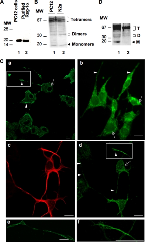
Reg-1α is expressed in PC12 and N2a cells as well as in embryonic hippocampal neurons (E17.5; 2 days in vitro). A, total cell protein extracts (20 μg) from differentiated PC12 cells grown in the presence of 50 ng/ml NGF for 48 h were separated by 15% SDS-PAGE, and Western blot analysis was performed using a rabbit polyclonal anti-Reg-1α antibody. Molecular weight markers (MW) are indicated on the left. Lane 1, Reg-1α in PC12 cells has an apparent molecular mass of ∼20 kDa; lane 2, the specificity of the anti-Reg-1α antibody was tested using purified human recombinant Reg-1α, which shows an apparent molecular mass of 18 kDa. B, PC12 (lane 1) and N2a (lane 2) cell extracts were separated by 12.5% SDS-PAGE, and Western blot analysis using the rabbit polyclonal anti-Reg-1α antibody shows three clusters of Reg-1α expression with molecular masses of ∼70, 35, and 20 kDa (tetramers, dimers, and monomers of Reg-1α). Molecular weight markers (MW) are indicated on the left. C, immunofluorescence analysis in differentiated PC12 (panels a and e), N2a (panel b) cells, and hippocampal neurons (panels c, d, and f), which express β3-tubulin (panel c), show that Reg-1α is mainly localized at the plasma membrane (panels a, b, and d, arrowheads), particularly in growth cones (panels e and f; higher magnifications of the insets in panels a and d) and in the perinuclear region (panels a, b, and d, arrows). Images were visualized by confocal microscopy (Z-projections of three confocal optical sections; intervals, 0.6 μm). Scale bars, 10 μm. D, immunoblot analysis of membrane (lane 1) and cytoplasm (lane 2) fractions from differentiated PC12 cells using the polyclonal anti-Reg-1α antibody. Tetramers (T), dimers (D), and monomers (M) of Reg-1α are indicated. Molecular weight markers (MW) are indicated on the left.

