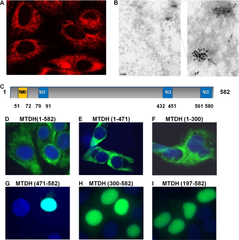
FIGURE 1.
MTDH is primarily cytoplasmic in cancer cell lines. A and B, subcellular distribution of MTDH and MTDH fragments was detected in Hec50 cells by confocal microscopy (A) and electron microscope (B) using a rabbit antibody against MTDH (amino acids 315–461). C, schematic representation of MTDH highlighting the NLS peptides. Orange box, transmembrane domain (TMD). Blue boxes, three NLS regions. D–I, distribution of exogenous FLAG-tagged full-length MTDH (D), MTDH C-terminal truncation mutants (E and F), and MTDH N-terminal truncation mutants (G–I) was detected with FLAG antibody. Nuclei were stained with DAPI.

