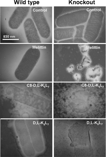
FIGURE 2.
Electron microscopy images of S. typhimurium SL1344 WT and phoPQ::Tn10 knock-out mutant incubated with peptides. Bacteria were incubated with either melittin (mutant MIC value), d,l-K5L7 (mutant MIC value), or C8-d,l-K5L7 (WT MIC value). The reason for selecting these MICs is detailed under “Experimental Procedures.” A drop of each sample was deposited onto a carbon-coated grid and negatively stained with phosphotungstic acid (2%, pH 6.8), and the grids were examined by electron microscopy.

