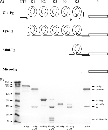
FIGURE 1.
Plasminogen variants. A, secondary structures of Pg variants displaying the kringle and protease domains of Glu-, Lys-, Mini-, and Micro-Pg. The asterisks (*) represent Pn cleavage sites between Lys-77 and Lys-78 (8, 30) and between Arg-530 and Lys-531 (31). The open arrow represents the elastase cleavage site between Val-441 and Val-442 (16). The solid inverted arrow indicates the tPA or uPA cleavage site between Arg-561 and Val-562 (1, 32, 33). NTP, NH2-terminal peptide; K1 through K5, kringle domains 1 through 5; P, protease domain. B, SDS-PAGE analysis of the isolated Pg variants under reducing conditions before and after uPA treatment to convert them to their respective Pn forms. Molecular weight markers are labeled on the left. Pn, obtained by uPA activation of Glu-Pg, was included as a control. The identities of the protein bands are labeled on the right. Lys-Pn-HC, Lys-Pn heavy chain; LC, light chain; Mini-Pn-Apep, Mini-Pn activation peptide.

