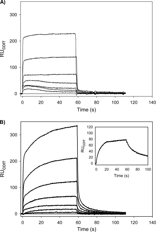
FIGURE 5.
SPR analysis of the interaction of Mini-Pg with Fn in the absence or presence of FPR-tPA. Mini-Pg, at varying concentrations (0, 0.31, 0.63, 1.25, 2.5, 5, 10, and 20 μm), was injected for about 60 s into a SA chip flow cell containing immobilized biotin-Fn in the absence (A) or presence (B) of 25 nm FPR-tPA to measure association. Cells were then washed with HBSC-Tween 20 to monitor dissociation. Between injections, the flow cell was regenerated with 1 m NaCl containing 20 mm ϵ-ACA in HBSC-Tween 20. In the absence of tPA (panel A), the Kd value determined was 31.3 μm. With FPR-tPA co-injection (panel B), Kd values for the high and low affinity binding sites determined using a two-site model were 35.8 and 131 μm, respectively. The inset in panel B displays the slow association and dissociation of the interaction of 25 nm FPR-tPA with immobilized Fn.

