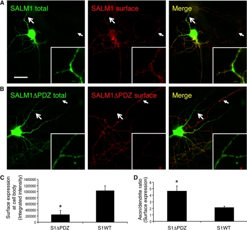
FIGURE 2.
SALM1 surface expression is mediated by the PDZ-binding domain. A, primary hippocampal neurons were transfected with Myc-SALM1 or Myc-SALM1ΔPDZ at DIV 12, and immunocytochemistry was performed 48 h later. Transfected SALM1 is expressed throughout the neuron and traffics readily to the surface of axons, dendrites, and the soma as detected by an N-terminal SALM1 (anti-S1NT) antibody (surface). Intracellular SALM1 was detected using an anti-Myc antibody (total). B, transfected SALM1ΔPDZ is expressed throughout the neuron, although the surface expression at somatodendritic sites is significantly reduced. The large arrows indicate dendritic arbors (magnified in insets), whereas the small arrows indicate axonal processes. C, quantification of SALM1 and SALM1ΔPDZ surface expression (integrated intensity) at the cell body. Deletion of the PDZ-BD significantly reduces somatic surface expression of SALM1ΔPDZ (241,944 ± 140,318, n = 6; *, p < 0.05, Student's t test) compared with SALM1 (1,029,640 ± 155,449, n = 7). D, quantification of the surface expression ratio (integrated intensity) of axons and dendrites of SALM1 (2.08 ± 0.27, n = 7) and SALM1ΔPDZ (4.61 ± 0.79, n = 6; *, p < 0.05, Student's t test) transfected neurons indicates that SALM1ΔPDZ has a significantly higher axon/dendrite ratio than SALM1. Values are shown as mean ± S.E. (error bars). Scale bar, 20 μm.

