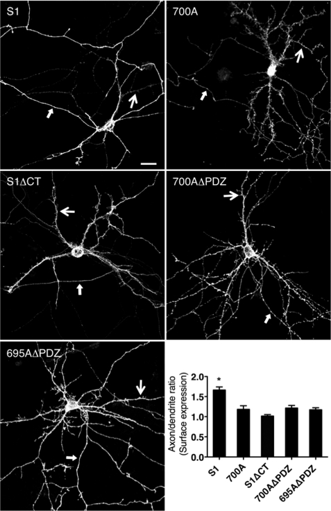
FIGURE 6.
Loss of dileucine motif enhances SALM1 dendritic localization. Primary hippocampal neurons were transfected with Myc-SALM1, Myc-SALM1 L700A, Myc-SALM1ΔCT, Myc-SALM1 L700AΔPDZ, or Myc-SALM1 D695AΔPDZ at DIV 12 (compare with Myc-SALM1ΔPDZ in Fig. 2). Surface immunocytochemistry was performed 48 h later using an N-terminal SALM1 antibody. Confocal images of neurons transfected with Myc-SALM1 L700A show an increased complexity of the dendritic arbor, with highly prominent irregular spines and filopodia. Quantification of the surface expression (integrated intensity) of the SALM1 constructs indicates that SALM1 has a higher axon/dendrite ratio than SALM1 L700A, SALM1ΔCT, SALM1 L700AΔPDZ, and SALM1 D695AΔPDZ. Results are mean ± S.E. (error bars) (S1, 1.68 ± 0.087, n = 8; 700A, 1.19 ± 0.10, n = 8; S1ΔCT, 1.00 ± 0.04, n = 10; 700AΔPDZ, 1.26 ± 0.07, n = 8; 695AΔPDZ, 1.22 ± 0.05, n = 6; one-way ANOVA; *, p < 0.05). Scale bar, 20 μm; large arrows indicate a dendrite, and small arrows indicate an axon.

