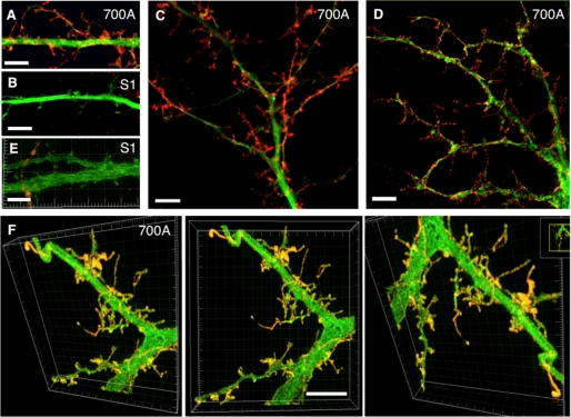
FIGURE 7.
Enhanced SALM1 surface expression increases filopodia and dendritic process formation; high resolution light microscopy. Shown are examples of dendrites from hippocampal neurons transfected with Myc-SALM1 or Myc-SALM1 L700A and GFP (green) at DIV 12 and surface-stained with an N-terminal SALM1 antibody (S1NT) 48 h later. Images were taken using laser confocal microscopy with a 1.46-nm objective lens (Zeiss) (A–C), wide field restoration microscopy (DeltaVision) (D), and three-dimensional structured illumination microscope superresolution imaging (DeltaVision) (E and F; profiles are shown from various angles in the same three-dimensional reconstruction) and show SALM1 surface puncta (red, anti-S1NT). High resolution microscopy illustrates the length and shape of the filopodia and processes that form as a result of L700A expression. L700A-transfected neurons (A and C–F) have larger and more irregular processes on the surface of their dendrites compared with SALM1 WT (B and E). Scale bars, 5 μm.

