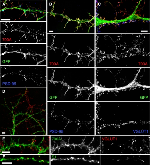
FIGURE 8.
SALM1 L700A localization studies show processes lacking synapses. Representative immunofluorescent images of hippocampal neurons transfected at DIV 12 and immunostained after 48 h. Wide field restoration microscopy (DeltaVision) (A) and confocal microscopy (B) show surface expression of SALM1 L700A (red) and endogenous PSD-95 (blue). The postsynaptic marker PSD-95 (blue) is present in the filopodia that form as a result of L700A expression (red, surface, S1NT antibody; green, GFP). On the other hand, L700A does not necessarily colocalize with the presynaptic marker, VGLUT1 (blue), that is present along the dendrite, and VGLUT1 does not colocalize with the longer filopodia (C). Total staining for L700A (D–E) or WT SALM1 (F) (green, total, S1NT antibody) and endogenous VGLUT1 (red) shows that L700A is present throughout the dendrite and filopodia, whereas VGLUT1 (red) mainly localizes along the dendrite shaft, at the base of the filopodia, and not at their tips. Scale bars, 5 μm.

