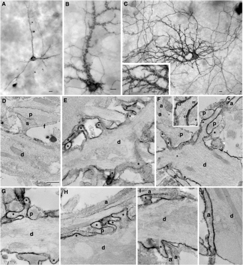
FIGURE 9.
Enhanced SALM1 surface expression increases dendritic arborization; electron microscopy. Shown is immunoperoxidase/DAB surface labeling of transfected SALM WT (A) or L700A (B–J) with light microscopy (A–C and inset) and EM (D–J) using an N-terminal SALM1 antibody. WT shows relatively smooth dendrites with a few labeled processes and low surface labeling (axons also labeled but not shown). In contrast, light microscopy of L700A shows “rough” dendrites covered in processes and densely labeled. Labeling in axons is also extensive. With EM, compare the relatively smooth, unlabeled surface of dendrites (d; thick, with tapering sides and containing bundles of microtubules, elongate mitochondria, and endosomes and other tubulovesicular structures) from untransfected neurons (D and J) with the irregular and process-laden (asterisks) labeled surface of dendrites of transfected neurons (E–I). Examples of presynaptic terminals (containing a cluster of synaptic vesicles) forming synapses include one on a normal spine (s; plus filopodium, f) in D, whereas another presynaptic terminal makes synapses near the base of the long process in F (synaptic vesicles (v) and synaptic clefts (cl) are labeled in the inset); the latter process appears to have at least one microtubule (m in inset) and is probably a developing dendritic branch. Other presynaptic terminals appear to be making synaptic contact with the dendrite shafts or the base of the adjacent process (a cleft is not distinct in these sections; left p in F and in G and H). Axons (a; thin, with roughly straight, parallel sides and containing microtubules) from untransfected (unlabeled axons in F, H, and I) or transfected (labeled axon in J) neurons often form in bundles on the surface of dendrites (H and I) as seen in normal cultures (14). Unmarked neurites in the micrographs may be dendrites or axons. Scale bars, 10 μm for light microscopy (A–C) and 500 nm for EM (D–J; 100 nm for inset in F) micrographs.

