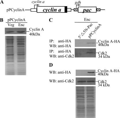
FIGURE 8.
Interaction between Cyclin A and Cdk2. A, diagrams of the 5′Δ5N-Pac and pPCyclinA plasmids. The pac gene (open box) is under control of the 5′- and 3′-flanking regions of the gdh gene (striated box). The cyclin a gene is under control of its own 5′-flanking region (open box) and the 3′-flanking region of the ran gene (dotted box). The filled black box indicates the coding sequence of the HA epitope tag. B, cyclin A protein levels in pPCyclinA stable transfectants. The pPCyclinA stable transfectants were cultured in growth (Veg, vegetative growth) or encystation (Enc, encystation) medium for 24 h and then subjected to SDS-PAGE and Western blot (WB). HA-tagged Cyclin A proteins were detected using an anti-HA antibody by Western blot analysis. Equal amounts of protein loading were confirmed by SDS-PAGE and Coomassie Blue staining. C, interaction between Cyclin A and Cdk2 detected by co-immunoprecipitation assays. The 5′Δ5N-Pac and pPCyclinA stable transfectants were cultured in encystation medium for 24 h. Proteins from cell lysates were immunoprecipitated using anti-HA antibody conjugated to beads. The precipitates were analyzed by Western blot with anti-HA or anti-Cdk2 antibody as indicated. D, expression of HA-tagged Cyclin A and Cdk2 proteins in whole cell extracts. The 5′Δ5N-Pac and pPCyclinA stable transfectants were cultured in encystation for 24 h (Enc) and then subjected to Western blot analysis. The blot was probed by anti-HA and anti-Cdk2 antibody. Equal amounts of protein loading were confirmed by SDS-PAGE and Coomassie Blue staining.

