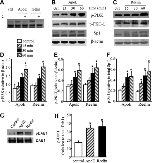
FIGURE 2.
Reelin and apoE3 increase Dab1, PI3K, PKCζ, and Sp1 phosphorylation. A, mouse macrophages were treated with 3 μg/ml apoE3, 2 μg/ml reelin, or culture medium alone as a control (ctrl) for 30 min. The lysates were then incubated with or without λ-phosphatase (λ). The Sp1 protein level was determined by Western blot analysis. B–F, mouse macrophages were treated with 3 μg/ml apoE3, 2 μg/ml reelin, or culture medium alone as a control for the indicated time periods. The levels of Sp1 and phosphorylated PI3K (p-PI3K) and PKCζ (p-PKCζ) were determined by Western blot analysis (B and C) and quantitated relative to β-actin (D and E). The level of phosphorylated Sp1 (top band for Sp1 in B and C) was quantitated relative to the level of total Sp1 (the sum of the top and bottom bands) and are shown in F. G and H, macrophages were treated with 3 μg/ml apoE3, 2 μg/ml reelin, or culture medium alone (control) for 1 h. Dab1 was immunoprecipitated. G, Western blot analysis was performed to detect the protein level of total Dab1 and phosphorylated Dab1 (pDab1) in the precipitant. H, the level of p-Dab1 was quantitated relative to the level of Dab1. *, p < 0.05 compared with controls.

