FIGURE 4.
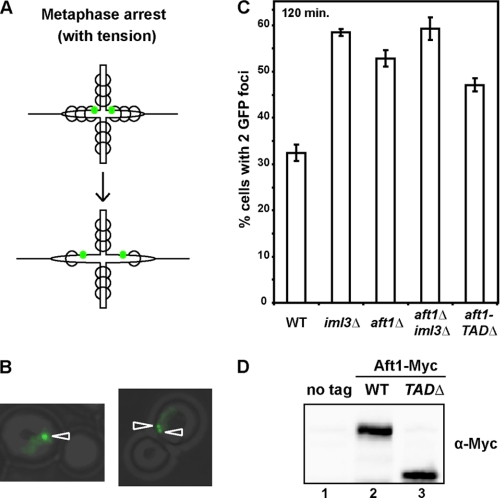
aft1Δ mutants exhibit pericentromeric cohesion defects in mitosis. A, schematic showing the separation of GFP-marked chromosomes 2.4 kb to the right of CEN4 in a metaphase arrest while maintaining microtubule tension in a strain with adequate pericentric cohesin (top panel) and decreased pericentric cohesin (bottom panel). Spindle axis is along the horizontal plane. GFP is represented by green dots. Cohesin is represented by circles. B, representative images of cells at 120 min after release from G1 showing one or two GFP foci (left and right panel, respectively). C, sister centromeres are more frequently separated in aft1Δ, iml3Δ, and aft1-TADΔ mutants. Wild-type (WT), aft1Δ, iml3Δ, and aft1-TADΔ cells that have GFP-labeled chromosome 2.4 kb to the right of CEN4 and MET-CDC20 were arrested in G1 with α-factor and released into media containing methionine to induce a metaphase arrest while maintaining tension between sister chromatids. Metaphase arrest was confirmed by bud count, and the frequency of GFP separation was assayed by microscopy. Results are the mean of three experiments in which 200 cells were scored. D, removal of the transactivation domain does not impact protein levels. Western analysis of whole-cell extract from wild-type (no tag), Aft1-Myc, and aft1-TADΔ-Myc cells.

