FIGURE 1.
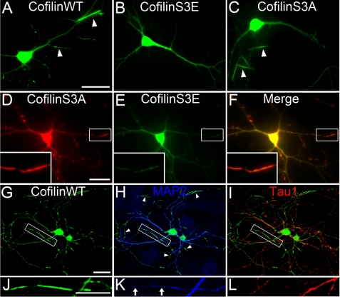
Cofilin rods form in distal dendrites. A, representative image shows cofilin rods (indicated by white arrowheads) in neurons transfected with wild-type GFP-cofilin (7 days in vitro transfection and 9 days in vitro imaging). B and C, overexpression of GFP-cofilin S3E did not induce rod formation whereas transfection of GFP-cofilin S3A did induce rod formation. D–F, coexpression of RFP-cofilin S3A and GFP-cofilin S3E resulted in rod formation with RFP-cofilin S3A concentrating in rods and GFP-cofilin S3E largely absent in rods. Inset in left corner is the enlarged view of a cofilin rod. Note the presence of RFP-cofilin S3A but not GFP-cofilin S3E in rod region. G–I, immunostaining with dendritic marker MAP2 (blue) and axon marker tau1 (red) showed that the majority of cofilin rods were formed in distal dendrites and less in axons. J–L, enlarged view shows cofilin rods in dendrites versus axons. Note that MAP2 staining is significantly reduced in rod areas, suggesting a possible destruction of microtubule integrity in rod regions. Scale bars, 20 μm.

