FIGURE 2.
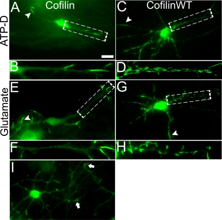
Neurotoxic stimulation induces cofilin rod formation. A, hippocampal neurons were immunostained with cofilin-specific antibodies after being stimulated with ATP depletion medium (10 mm NaN3 and 6 mm 2-deoxyglucose, 30 min). After stimulation, endogenous cofilin formed rod structures. B, enlarged view of rods in A are shown. C and D, neurons were first transfected with cofilin-GFP and then stimulated with ATP depletion medium. Note severe rod formation in these cofilin-transfected neurons. E and F, glutamate (200 μm, 30 min) stimulation induced endogenous cofilin rod formation. G and H, neurons first transfected with cofilin-GFP and then stimulated with glutamate also showed severe rods. I, cofilin immunostaining affected nontransfected and nonstimulated control neurons. Arrows point to growth cones with accumulated cofilin signal. Scale bar, 10 μm.

