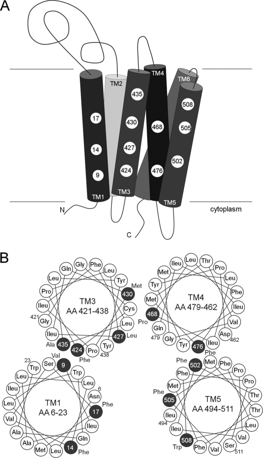
FIGURE 10.
Model of the YidC hydrophobic protein binding platform. A, the major contact sites in TM1, 3, 4, and 5 are depicted by a numbered circle referring to the amino acid residue position. B, helical wheel projection to illustrate the substrate contact sites of the YidC TM regions. The major contact sites are highlighted as dark circles with the respective position. Both ends of each TM segment are labeled with the position number outside the circle. Because TM2 and TM6 showed no strong contacts, they are not included in the model.

